Relation between Cardiac Output and Peak Plasma Propofol Concentration with Variable-rate Infusion on Same Dose
Masui K, MD, Kazama T, MD , Kira M, DDS. National Defense Medical College, Tokorozawa, Saitama, Japan
Background
Cardiac output (CO) affects plasma concentrations of intravenous drugs administered continuously. Kurita et al (Anesthesiology 2002; 96:1498) reported higher CO decreased plasma concentration of propofol than lower output at a same rate infusion. However, effect of CO on plasma concentration during the initial infusion of propofol had not been examined. The goal of this study was to examine whether or not infusion rate and CO affected the plasma concentration during the induction of anesthesia.
Methods
Fifty patients undergoing elective surgery were included. Exclusion criteria were patients younger than 20 yr, older than 75 yr, ASA physical status ≥III, pregnancy, body mass index ≥30, and significant heart, hepatic or renal impairment.
Patients were given no premedication. After entering the operating room, a 18 or 20 gauge forearm venous catheter for propofol infusion, and a 22 gauge contralateral radial artery catheter for taking blood samples were placed. Before the induction of anesthesia, CO was determined (preCO) using the DDG-3300 pulse dye densitometer (Nihon Kohden, Japan), and the CO value was used to calibrate PulseCO system (LiDCO, UK). Infusion rate of propofol was randomly assigned to 160, 80, 40, 20, or 10 mg/kg/hr (160, 80, 40, 20, or 10 group, respectively,). Propofol 1.2 mg/kg was administered via the forearm venous catheter directly. Arterial blood samples were taken every 5 sec during the first one minute, and every 10 sec before the end of propofol infusion until 90 seconds after the end of the propofol infusion. CO was measured with PulseCO system for 10 seconds just after the last blood sample taken, and the mean of these values was used for analysis (postCO). Arterial plasma concentrations (Cp) of propofol were determined by high-performance liquid chromatography with fluorescence detection. Peak Cp was defined as the highest concentration detected and time to peak was defined as the time at which that concentration was measured.
Tukey and Steel-Dwass method were used for multiple comparisons among the groups. The correlation analysis was performed between cardiac output and peak Cp in each group. A P value of 0.05 was considered significant. Data expressed mean±SD or median [25percentile - 75percentile].
Result
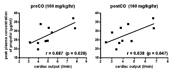
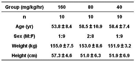
Differences in age, sex, weight, and height were not significant among the groups (not shown). Significant positive correlations between preCO and peak Cp (r=0.678), and between postCO and peak Cp (r=0.638) were observed in 160 group. Significant negative correlation between preCO and peakCp (r=0.701) was also observed, however there was no correlation between CO and peak Cp in the other groups. Times to peak were 45 [38-48], 80 [70-80], 130 [115-140], 225 [213-240] and 430 [423-438] sec in 160, 80, 40, 20 and 10 groups, respectively. Differences between all pair among the groups in time to peak were significant.
Conclusions
High CO increased peak Cp at 160 mg/kg/hr of propofol. On the contrary, high CO decreased peak Cp at 20 mg/kg/hr. Short peak time may affect the correlation between CO and peak Cp.
 |
Demographic Data
 |